Welcome to the second issue of Volume 3 of PLANE WAVE, Verasonics’ Newsletter through which we share information about new products and technologies, emerging applications, conferences, training opportunities, and collaborations with researchers in ultrasound and ultrasonic technologies. We hope you find these newsletters informative and interesting, and welcome your suggestions for future topics.
New Products and Applications ▪ Technology Information ▪ Research ▪ Conferences and Training
Verasonics Provides Outstanding Research Ultrasound Capabilities with the Vantage High Frequency Research Ultrasound Platform
High-frequency ultrasound imaging above 15 MHz has become a highly innovative research field. Although human clinical applications are still rare, pre-clinical high-frequency ultrasound has become a rapidly emerging discipline due to new technological advances in high-frequency arrays and signal processing.
Verasonics is proud to support exciting research developments in high-frequency ultrasound by providing researchers around the world with cutting-edge ultrasound technology through its Vantage platform. Verasonics’ Vantage systems deliver exceptional flexibility, ultra-fast frame rates and high-performance imaging, Doppler and focused ultrasound capabilities to pre-clinical ultrasound research. With high-frequency configurations and a set of four high-frequency transducers available on the Vantage 64LE™, Vantage 128™ and Vantage 256™ systems, Verasonics now offers imaging capabilities above 30 MHz. Figure 1 shows two images that were acquired using an L38-22v CMUT linear array transducer on a Vantage 256™ system. The images demonstrate the excellent spatial and contrast resolution that the Vantage system is capable of providing.

Figure 1 Examples of high-frequency-ultrasound images acquired using a L38-22v CMUT linear array of a) a ulnar artery and b) mouse kidney.
This superior flexibility of the Vantage Systems is enabled by the ability to adjust the ADC sample rate and to program an analog High-Pass Filter in addition to the conventional anti-aliasing Low Pass Filter. A flexible acquisition sequence control permits the user to choose between several different high-frequency acquisition approaches, depending on tradeoffs between frame rate, data bandwidth, and SNR. Inter-leaved sampling is possible using two consecutive acquisitions because the ADC integration window is short enough for 125 MHz sampling. Bandwidth sampling approaches are used when a single acquisition is required. Example imaging programs are provided with the system software for 4x sampling (where x is the transducer center frequency), 4/3x sampling, 8/3x sampling, inter-leaved acquisitions, and for simple data collection and transfer at any digitization frequency available. For more information about these signal processing techniques please read Verasonics’ Bandwidth Sampling white paper.
High-Frequency Ultrasound Case Studies with the Vantage System
Recently, Dr. Daniel Rohrbach joined Verasonics and will support new software developments for high-frequency applications of the Vantage systems. Prior to joining Verasonics, Dr. Rohrbach worked at the Lizzi Center for Biomedical Engineering at the Riverside Research Institute as a principal investigator where he collaborated with some Verasonics’ customers. These customers included Dr. Jeffrey A. Ketterling from the Lizzi Center for Biomedical Engineering at the Riverside Research Institute and Dr. Ronald H. Silverman from the Department of Ophthalmology at the Harkness Eye Institute Columbia University in New York City. Dr. Ketterling and Dr. Silverman are both considered leading researchers that have successfully used the high-frequency-ultrasound capabilities offered with the Vantage system for research.
Dr. Ketterling and team uses vector-flow analysis methods to study blood-flow patterns in the mouse embryo[1]. Imaging blood flow in the hearts of embryonic mice, in utero, can be challenging due to the small size of the heart and rapid heart rate. High-frequency ultrasound systems using single-element and linear-array transducers have been used for many years to obtain local Doppler-flow estimates in embryos. However, these systems are limited and unable to estimate the instantaneous flow at high temporal and spatial resolution over a large area. Additionally, they are not able to portray the complex nature of blood-flow patterns in the heart.
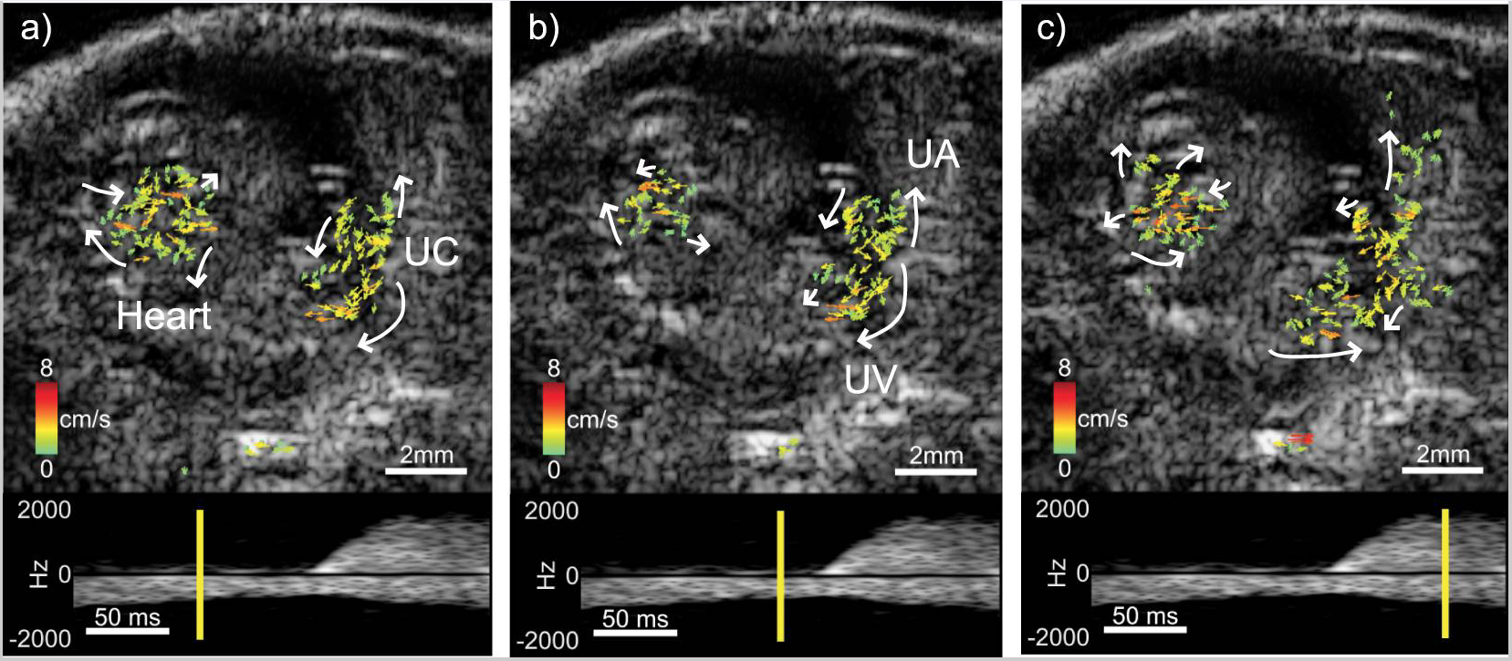
Together with collaborators at the NYU School of Medicine and the University of Waterloo, the group and Dr. Ketterling performed initial experiments applying plane-wave, high-speed imaging and vector-flow estimate approaches to blood flow in mouse embryos. An 18-MHz, L22-14v linear array was used to acquire plane-wave data from a 16.5 day old embryo at an absolute frame rate of 20 kHz using a set of five fixed transmission angles. After beamforming, vector-flow processing and image compounding, the effective frame rate was 4 kHz. The vector-flow analysis revealed the complex nature of the blood flow patterns in the embryo and umbilical cord. Figure 2 demonstrates different phases of the cardiac cycle: (a) early systolic phase, (b) late diastole to early systole, and (c) the flow in the heart chambers while the umbilical artery reveals an increased flow after the earlier heart contractions. These initial results indicate that high-frequency, plane-wave imaging has the potential to simultaneously resolve complex cardiac motion and blood-flow patterns throughout a full image frame at sub-millisecond temporal resolution.
[1] Ketterling JA, Aristizábal O, Yiu BYS, Turnbull DH, Phoon CKL, Yu ACH, Silverman RH. High-speed, high-frequency ultrasound, in utero vector-flow imaging of mouse embryos. Sci Rep. 2017 Nov 30;7(1):16658.

Figure 2 Vector-flow imaging of an mouse embryo heart using the Vantage system and the L22-14v transducer.
In another case study under IRB approval, Dr. Silverman and Dr. Raksha Urs (Harkness Eye Institute, Columbia University) used the Vantage system with an L22-14v linear array probe to study blood flow in the eye[1],[2]. Plane-wave imaging methods are particularly advantageous in ophthalmology because acoustic intensity may be significantly lower than it is by using conventional Doppler methods.
Scans centered on the optic nerve head are typically acquired for 3 seconds, allowing capture of several cardiac cycles. In this example, two angled plane-waves were transmitted at ±9 degrees at a 6 kHz PRF after compounding. In Figure 3, the leftmost image is the structural B-scan showing the mostly anechoic vitreous (V), the vitreo-retinal interface (R), and the optic nerve (ON). The center image shows a directionally color-coded power Doppler image, derived from the same data. On the right is a spectrogram portraying flow velocities in the region of interest (box) in the power Doppler image capturing flow velocities from both the central retinal artery and vein.
[1] Urs R, Ketterling JA, Yu ACH, Lloyd HO, Yiu BYS, Silverman RH. Ultrasound imaging and measurement of choroidal blood flow. TVST 2018;7(5);1-8.
[2] Urs R, Ketterling JA, Silverman RH. Ultrafast ultrasound imaging of ocular anatomy and blood flow. Invest Ophthalmol Vis Sci. 2016:57(8):3810-6.

Figure 3 Scans of the optic nerve: (from left to right) B-Mode image, directionally color-coded power Doppler image, spectrogram portraying flow velocities
High Frequency Transducers Available for a Complete High Frequency Ultrasound Research Solution
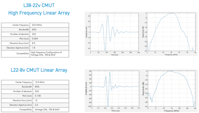
Verasonics brings the exceptional flexibility and performance of its Vantage research ultrasound systems to pre-clinical and small animal imaging. With high frequency, broadband transducers ranging up to 30 MHz center frequency with either single crystal or CMUT technologies, the Vantage system is now capable of providing the platform for many cutting-edge applications and techniques. These transducers, together with the Vantage system provide ideal ultrasonic configurations for scientists that are conducting pre-clinical research for new diagnostic and therapeutic techniques and products.
KOLO Medical Transducers
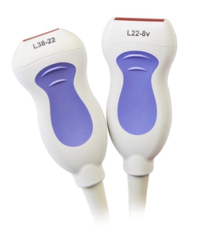
The CMUT transducers from Kolo Medical have 256 elements multiplexed into 128 channels and are compatible with the Vantage 64 LE™, Vantage 128™ and Vantage 256™systems. They provide superb performance for pre-clinical imaging, including mice and rats, and for other superficial imaging applications.


Vermon Transducers
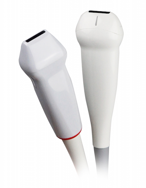
Vermon develops and manufactures state of the art single-crystal transducers for biomedical research. Verasonics offers two Vermon high frequency linear array transducers for pre-clinical ultrasound research. These transducers provide high-quality imaging and Doppler performance for small animals and for other superficial imaging applications.


Visit Us and Learn What’s New at these Upcoming Conferences:
Currently there are no upcoming conferences, however, please join us for our next Vantage customer training via live webinar, 1-4 February, 2021, 9 – 11 am PT.
Please contact [email protected] to register or click here for more information.

