Vantage Application Gallery
Microvascular Imaging
Vascular imaging of a rabbit kidney using high frame rate contrast enhanced acquisition and signal processing through ASAP with the L12-3v linear array. The lateral dimension of the image is 2.5cm. Frame rate is 750 frames per second.
Reference: Stanziola A et al., IEEE TMI, V37(8), pp 1847 – 1856, Aug. 2018
(title: ASAP: Super-Contrast Vasculature Imaging using Coherence Analysis and High Frame-Rate Contrast Enhanced Ultrasound)
Video courtesy of Professor Mengxing Tang, Imperial College London.
High Frame-Rate Imaging
High Frame Rate contrast echocardiography of a human volunteer’s heart using a P4-1 phased array transducer, acquired at 5500 frames per second.
Reference: Toulemonde MEG et al., JACC Cardiovascular Imaging, V11(6), pp 923-924 June 2018
(title: High Frame-Rate Contrast Echocardiography: In-Human Demonstration)
Video courtesy of Professor Mengxing Tang, Imperial College London.
High Frequency Imaging
B-mode imaging of a 1mm vein in the finger, acquired with a CMUT linear array transducer at 41 MHz. Notice the detail in the wall of the vein.
High Frequency Imaging
Venous valve leaflets in a vein in the arm, acquired with the KOLO L38-22v CMUT Linear Array transducer. This 30MHz image shows structure of the vein wall and blood flow through the valve with respiration.

Power Doppler
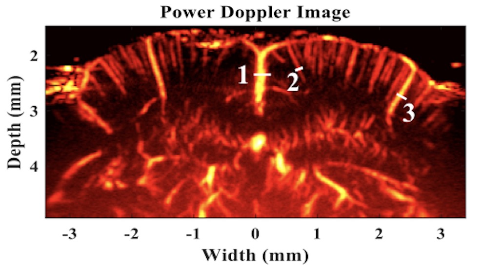
High resolution image of a mouse brain produced using ultrafast power Doppler acquisition sequences without micro bubbles. The image, showing vascularity within the hippocampus, was acquired using a Vantage 256 High Frequency configuration and a 40 MHz transducer from Visualsonics.
Image courtesy of Prof. Chih-Chung Huang, Department of Biomedical Engineering, National Cheng Kung University, Tainan, Taiwan
Pre-clinical Imaging
Hand-held, b-mode imaging of a mouse kidney, acquired at 30MHz using a wide-beam, spatial compounding script on a Vantage 128 High Frequency system with the L38-22v CMUT linear array.

HIFU and FUS Research
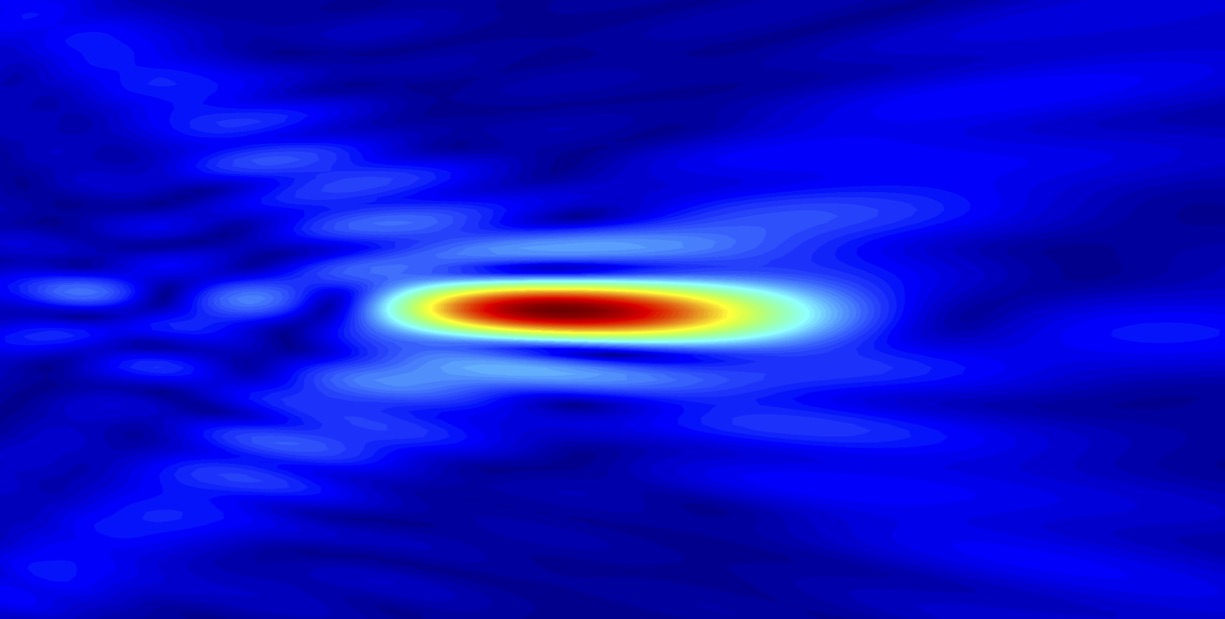
Focused Ultrasound delivers high intensity energy in very small areas, enabling research in therapeutic techniques that utilize thermal effects, cavitation or histotripsy. This image shows the focal spot of a HIFUPlex simulation.
For more information about HIFUPlex, please click here
Biometric Research
Finger print
This video was acquired with the finger immersed in water, and the transducer scan plane tangential to the finger surface. Vantage systems enable research and development in novel biometric techniques.
High Frame Rate
High frame rate contrast enhanced imaging and quantification of arterial flow and wall shear stress on an arterial (carotid) flow phantom with stenosis, using the L12-3v linear array.
Reference: Leow CH et al, Ultrasound in Med. & Biol., V44(1), pp. 134–152, 2018
(title: SPATIO-TEMPORAL FLOW AND WALL SHEAR STRESS MAPPING BASED ON INCOHERENT ENSEMBLE-CORRELATION OF ULTRAFAST CONTRAST ENHANCED ULTRASOUND IMAGES)
Video courtesy of Professor Mengxing Tang, Imperial College London.
High Frequency
This cross-sectional image clip of the wrist shows the median nerve structures. It was acquired on a Vantage high frequency system at 41 MHz using a CMUT linear array transducer.
Musculo-Skeletal Imaging
Flexor tendon in the finger, showing its co-axial structure and independent movement of the proximal and distal portions of the tendon. Images acquired with a 256-element CMUT linear array at 41 MHz on a Vantage 256 high frequency system.
Doppler Imaging
Vascular color Doppler clip obtained with the Verasonics L11-5v linear array transducer, using a wide-beam Doppler acquisition script without any post-processing.
The Vantage System is intended to be used for research and experimental uses only.
The Vantage System is not a diagnostic ultrasound device.
Images shown are for demonstration purposes only.
11335 NE 122nd Way, Suite 100
Kirkland WA 98034

