
Functional Ultrasound: Advancing Non-Invasive Functional Imaging Research
Functional Imaging Today
For years, neuroscientists, physicians and biomedical engineers have sought to better visualize changes in the brain as a result of normal activity and external stimuli. It is believed that functional imaging has the potential to allow quantitative anatomical, temporal and metabolic information to be incorporated into more accurate diagnosis, monitoring, response to medication, and neurosurgical treatment planning. Functional imaging or physiological imaging also can be used beyond the brain as a technique for detecting or measuring changes in metabolism, blood flow, chemical composition and absorption, such as with assessment of angiogenesis in tumors.
Various techniques have been explored with functional imaging, including functional magnetic resonance imaging (fMRI), positron emission tomography (PET), electroencephalography (EEG), magnetoencephalography (MEG) and near infrared spectroscopy (NIRS). Limited imaging resolution and sensitivity, as well as cost, inaccessibility, and practical difficulties have prevented widespread adoption of these techniques.
Research in Functional Ultrasound (fUS): The Promise of a Game-Changing Modality
Over the last decade a growing number of researchers have been studying ultrasound as a new tool for imaging brain function. Capitalizing on the real-time nature and high resolution of ultrasound, researchers have demonstrated that they can rapidly assess hemodynamics and vascular changes in the brain due to stimuli response. Additionally, fUS can be used to provide high quality imaging for identifying structures involved in cognitive processes. fUS is now being evaluated in the diagnosis of diseases such as Parkinson’s, Alzheimer’s, and epilepsy.
One of these researchers is Emilie Mace, PhD, at Friedrich Miescher Institute for Biomedical Research in Switzerland. She believes that “Ultrasound’s tremendous flexibility and accessibility, and high spatial resolution and sensitivity over other imaging modalities, make it an ideal imaging modality to perform functional activity mapping of the brain. In addition, ultrasound provides a real time and portable technique for awake, moving small animal studies.”
Dr. Mace and colleagues have pioneered techniques for fUS that utilize ultrafast, plane-wave imaging modes, with high frequency transducers, to measure blood flow changes in small vessels in the brains of rats. Her studies have demonstrated it is possible to identify vascular response in regions of brain activation following whisker stimulus, and visualize perfusion changes associated with epileptic seizures. (Functional Ultrasound Imaging of the Brain, published in Nature Methods in 2011)
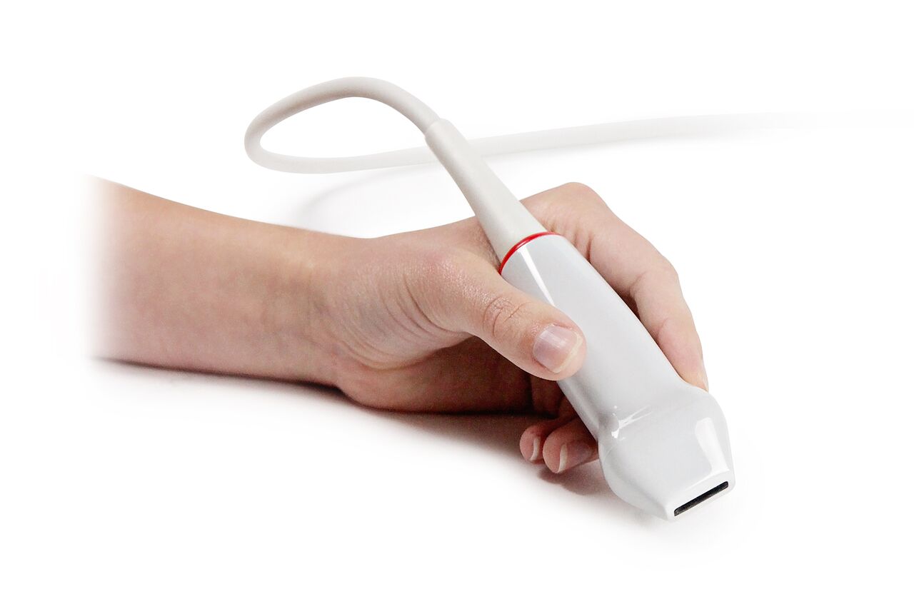 Dr. Mace uses a Verasonics Vantage 128™ research system, with a single crystal, 128-element L22-14v linear array transducer at a center frequency of 18.5 MHz. The Vantage system’s ability to acquire plane wave images at thousands of frames per second enables Dr. Mace to use advanced acquisition and signal processing techniques for her small animal studies. “Functional Ultrasound has great potential to advance neuroscience in the future. With high frame rates we can obtain high resolution and sensitivity to gain valuable insight into the way the brain functions,” said Dr. Mace. “The flexibility of the Vantage system’s architecture to easily program exploratory sequences for new algorithms has been extremely important to advance our research.”
Expert researcher, Paul Dayton, PhD, specializes in this rapidly evolving field, however his team at the Dayton Lab in North Carolina explores non-invasive fUS and therapeutic techniques. Dr. Dayton is also professor at the joint biomedical engineering department of University of North Carolina and North Carolina State University. His lab focuses on contrast-enhanced ultrasound pre-clinical research with imaging techniques including perfusion and molecular imaging, acoustic angiography, and contrast agents with phase change perfluorocarbons and monodisperse microbubbles.
In January 2017, Dr. Dayton working with collaborator Dr. Gianmarco Pinton published a paper in Theranostics entitled, “3-D Ultrasound Localization Microscopy for Identifying Microvascular Morphology Features of Tumor Angiogenesis at a Resolution Beyond the Diffraction Limit of Conventional Ultrasound.” Dr. Dayton and team studied the theory of ‘ultrasound localization microscopy’ by applying a combination of volume with quantitative analysis of microvascular morphology to assess angiogenesis in cancers. Ultrasound has traditionally been technically limited in microvascular imaging. This study used a Vantage research ultrasound system and demonstrated the ability to image complex microvascular patterns associated with tumor angiogenesis in-vivo at a resolution of tens of microns – substantially better than the diffraction limit of traditional clinical ultrasound, while using an 8 MHz clinical ultrasound probe. Results suggested that with continued development, ultrasound has the potential to detect biomarkers of cancer based on the microvascular ‘fingerprint’ of malignant angiogenesis rather than through imaging of blood flow dynamics or the tumor mass itself.
![]() Imaging was performed using the Vantage system with a L11-5 linear probe, using plane-wave imaging at a pulse repetition frequency of 500 Hz. The transmitted pulses were 1 cycle sinusoids at 4.5 MHz with a rarefactional pressure of 220 kPa (mechanical index = 0.1). This low mechanical index was chosen to minimize bubble destruction under high frame rate insonification. The transducer was mounted to a motorized precision motion stage synchronized with the imaging system for 3D scanning. Super resolution imaging was achieved with 8,000 images being acquired for each scan slice, and slice step size was 200 µm. All rats were also imaged with the super-harmonic imaging based acoustic angiography technique for comparison.
Imaging was performed using the Vantage system with a L11-5 linear probe, using plane-wave imaging at a pulse repetition frequency of 500 Hz. The transmitted pulses were 1 cycle sinusoids at 4.5 MHz with a rarefactional pressure of 220 kPa (mechanical index = 0.1). This low mechanical index was chosen to minimize bubble destruction under high frame rate insonification. The transducer was mounted to a motorized precision motion stage synchronized with the imaging system for 3D scanning. Super resolution imaging was achieved with 8,000 images being acquired for each scan slice, and slice step size was 200 µm. All rats were also imaged with the super-harmonic imaging based acoustic angiography technique for comparison.
“This study demonstrated an important breakthrough in functional imaging as we explore new approaches to understanding tumor angiogenesis,” said Dr. Dayton. “With ultrasound and contrast we have a low cost, flexible and less labor intensive technique in comparison to other modalities, such as MRI.”
Dr. Dayton added, “The Vantage ultrasound system played a critical role in this study and our ongoing research as a highly programmable, cutting edge ultrasound technology. It allows us to perform high quality contrast-enhanced imaging and acquire large quantities of data very fast, proving ‘ultrasound localization microscopy’ is a viable technique in the way tumors can be staged at a microvascular level.”
Introducing New Matrix Array Transducers for Volume Imaging: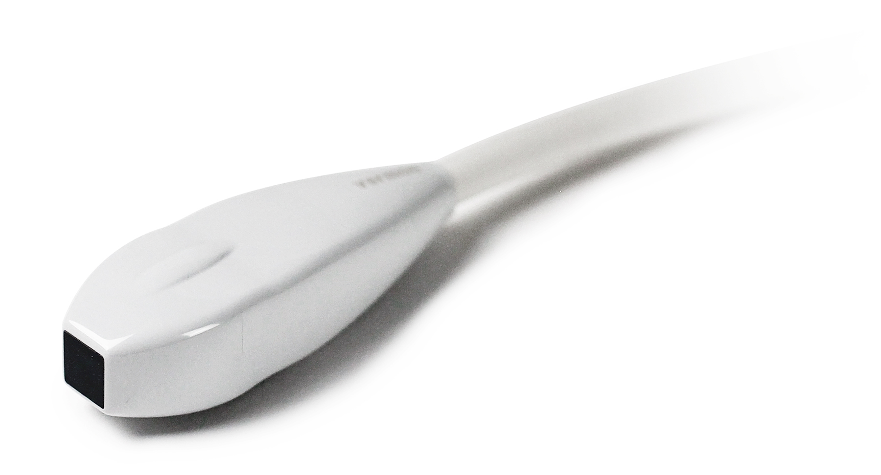
Verasonics is pleased to announce availability of two matrix array transducers with 1024 elements. Read more.